Category: Uncategorized

  • Caries Detection: Examining restorations, and when repair or replacement is required.

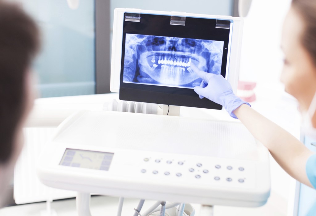
    Examining restorations:

    The presence of defective restorations or restorations with the clinical diagnosis of secondary caries is one of the most frequent problems encountered by general practitioners today. The diagnosis is inconsistent among dental practitioners and often does not rely on objective criteria. If in doubt, most general dental practitioners choose replacement as opposed to options of non-surgical treatment, including systematic restoration monitoring. Restoration replacement is especially common for restorations not originally placed by the evaluating practitioner. A dental practice-based research study involving 197 clinicians from the USA and Scandinavian countries, and close to 10,000 restorations, indicated that practitioners chose replacement over repair of defective dental restorations in over 75% of cases. The same study confirmed that practitioners who did not place the original restoration were more likely to replace it than practitioners who did.

    In summary, replacement of restorations constitutes over 50 per cent of the work performed by general dental practitioners in their practices and it has contributed to the perpetuation of the “Repeat Restoration Cycle”. Consequently, the diagnostic finding ‘defective’ for an existing restoration is a critical step in treatment planning and it invariably affects the longevity of the restored tooth.

    Secondary caries and staining of the margins of existing restorations are the most commons reasons for restoration replacement in permanent and primary teeth. Without objective criteria, it is difficult to differentiate secondary caries from marginal staining clinically. Despite the fact that some studies have associated microleakage with secondary carious lesion formation, the majority of the evidence demonstrates no relationship between the development of secondary carious lesions and the size of the leakage or gap alongside a restoration, except in cases in which the crevice exceeds 400 µm. Although the criteria for the diagnosis of a defective restoration may be based solely on visual and tactile examination, the subsequent management plan for this restored tooth should be based on the caries risk assessment of the patient as well.

    When is repair or replacement required?

    Laboratory and clinical studies have shown that removal of the existing restoration will remove significant healthy tooth structure, subsequently resulting in larger dental restorations. The removal of existing restorations may also cause additional stress on the tooth, with possible pulp reaction to thermal, chemical, bacterial, or mechanical stimuli, depending on the size and depth of the existing restoration.

    Therefore, the decision to replace existing restorations should be taken cautiously, as it may significantly affect the remaining tooth structure and, consequently, impact the longevity of future restorations and the lifespan of the tooth. Studies have demonstrated that replacing an existing restoration will not necessarily guarantee that the new restoration will surpass the clinical performance of other alternative treatments such as repair, sealing or monitoring.

    Long-term clinical studies have also shown that when alternative treatments fail, the failure usually takes place within 24 months. When the clinician is evaluating an existing restoration with one or more localized clinical features that deviate from ideal and the restoration is considered defective, the clinician should assess whether the tooth in question will truly benefit from a new restoration. When the practitioner is faced with a borderline situation, the patient’s past dental history and current caries risk status, and the best treatment for the tooth in question should be considered. If the practitioner is unsure whether the defective area can be removed by polishing or by sealing the affected area, another conservative and predictable approach would be to repair the restoration by removing the deteriorated area and re-restoring this area only.

    Generally, replacement should only take place if the practitioner cannot properly manage the defective areas without removing the entire restoration, or if there are pulpal symptoms.

    How successful are repaired restorations?

    Minimal Intervention Dentistry aimed to limit unnecessary removal of healthy tooth structure, and repair of defective restorations is one of its strategies. Although the repair of resin composite restorations has been investigated extensively and found successful, dental practitioners do not routinely consider this treatment option in the management of defective restorations.

    Although considered a long lasting treatment by the schools teaching this practice, a practice-based research study showed that only practitioners who practiced in non-fee service settings, practitioners with fewer years since graduation from dental school, and practitioners who assessed caries risk, chose preventive treatment options more often than replacement when assessing defective restorations The preference for replacement of restorations may be the result of a complex interplay between the lack of clear standards for replacing restorations and lack of an existing reimbursement for these treatments. That same study reported that general practitioners would most likely intervene surgically in a defective resin composite restoration but not in a defective amalgam restoration.

    So far, prospective studies have shown that repaired restorations in permanent teeth have the same or increased longevity as restorations that were replaced completely. Repair treatment remained stable over a 7-year observation period. Additionally, the reason that repaired restorations may even outlast those that were replaced probably relates to the fact that most of the restoration’s original form is kept intact, limiting the introduction of new elements that can affect the success of the restoration. When other restoration stress factors are considered, such as stress on the tooth, post-operative sensitivity, and re-exposure of the dentinal tubules with possible pulpal reactions to thermal or mechanical stimulus, damage to the adjacent tooth and the possibility of more complex restorations, it makes perfect sense to pursue the repair of defective restorations as a predictable and conservative approach to preserving tooth structure. A recent overview regarding restoration margins concludes that margin defects, without visible evidence of soft dentin on the wall or base of the defect, should be monitored, repaired or resealed, in lieu of total restoration replacement.

    Besides being a successful treatment, restoration repair is also practical. Defective restorations can be repaired more quickly and with lower operational costs than replacement. Therefore, repaired restorations could present a reduction in patient and/or the third party payers’ expenses which would potentially increase the number of individuals who could afford dental care. The cost of care and oral health are severely impacted by the replacement of existing restorations. Examining outcomes of alternative treatment to the replacement of restorations and establishing consistent criteria that will affect general practitioners’ treatment decisions is a critical issue that may profoundly change the over-treatment of existing restorations.

    In summary, dental practitioners should consider repairing truly defective restorations, an appropriate minimal invasive operative intervention worth pursuing.

  • Future of Dentistry and Dental Care

    Future of Dentistry and Dental Care

    Remember the biggest childhood nightmare?

    That you are sitting in from of many lights and someone is poking into your teeth with huge and frightening tools and then you get to know that you are in the dental clinic.

    The someone who was checking your mouth was, in reality, is a dentist and then he will ban your favorite eating items if you have found with any cavity.

    Moreover, to it, he will tell your mom to look whether you are brushing your teeth regularly or not and also order to brush twice. It is such a horrible nightmare which a child ever saw.

    But if you have any problem you have to go to the dentist to prevent your teeth from the further problem. Experts say and maybe you have heard in your childhood that your parents saying that “oral health is somehow connected to the overall health of the body”.

    It is not just a saying instead it is a truth.

    Now you don’t have to go to take various sessions to maintain your dental care. Dentistry has adopted various new technologies today, which can give instant and effective results.

    Dentistry has reached to this stage that now a person at the age of 80 can grow new teeth, don’t believe let’s see some of the modern technology that dentistry will adopt in the future to grow in coming time.

    Smart Toothbrush-

    At this period of technology our homes are filled with new technology and smart devices, why would not our bathroom?

    At first, it will feel a little bit strange to use a toothbrush with the sensor, but when you get habitual it will feel like a normal toothbrush.

    A smart electronic toothbrush makes sure that you are brushing your teeth in a right way.

    After some, it will become like an interesting game you are playing. This toothbrush combines with various sensors in the handle so that it can track how you are brushing your teeth. As you brush your teeth, it will show on the screen that how you are brushing your teeth, whether you are brushing particular teeth much or too little.

    Augmented Reality-

    As the “Pokemon GO” game become so famous and used worldwide, similarly, Augmented Reality also has the strength to transform the world especially in the world of healthcare.

    The Teledentistry-

    If you do not want to see a dentist, imagine how difficult it is for a child, a patient with a special need, or an old person in a sanatorium.

    Another problem is distance: people living in rural areas have little chance to enter the dentist, almost never have the possibility of choosing. With the spread of television, this may have changed a lot. It provides easier access to care for the patient, much cheaper for the patient – rather than expensive treatment, it turns to cheap preventive practices, and allow the patient to consult other unavailable medical professionals.

    Intra-Oral camera-

    The biggest inconvenience of a dentist’s chair is that if you cannot open your mouth, the doctor still does, but he cannot see what he wants to see, even in the famous dental mirror. Both the patient and the doctor suffer. Intra-Oral camera is a good solution to this exact problem. By the help of it, the doctor can easily see inside the mouth without doing much hard work.

  • Why Oral Hygiene is Important for Dental Health

    Why Oral Hygiene is Important for Dental Health

    Oral health is quite important since it does not only necessary to make you go with a flawless smile but also keeps you away from other diseases. Saying would not wrong that your mouth is a kind of window telling what is going inside your body. If you have been ignoring the dental health, you need to bring change to your habit.
    • Maintaining good oral hygiene is quite important as it plays a crucial role to keep you healthier and active longer. And it is not that much tough to keep as you just need to follow some easy steps.
    • Healthy teeth are not only important to have you a great smile or healthy teeth, but they also make it easy for you to eat and speak properly.
    • Saying would not wrong that good oral health is quite essential to your overall well-being. If you go for adopting healthy and good oral hygiene, it keeps you away from many health issues.
    • Good oral hygiene is important to keep you away from painful dental issues. It is all about your health adn overall body health and that is why it would not be right to ignore.

    What About The Relations Between Gum disease and health complications

    Some people avoid dental health issues thinking that it will not affect them a lot. According to Academy Of General Dentistry, there is a connection between gum disease and health complication. It indicates towards disease like stroke or heart disease. Apart from it, women having gum disease are higher risk of having pre-term or low bit.

    Women with gum disease also show higher incidences of pre-term, low birth-weight babies. Not only this, gum disease also indicates towards many other health issues like swollen gum, dry mouth, gum problems and so on.

    • Pancreatic cancer
    • Heart disease
    • Kidney disease
    • Diabetes
    • Leukemia
    • Oral cancer

    What You Can Do On Your Own

    • The fact cannot be denied that you need to visit the dentist if going through any sorts of dental issues. But you also need to know that what you can do on your own at home to keep your teeth healthy longer.
    •  Always brush twice in a day for at least two minutes, having fluoridated toothpaste. If you have swelling gums then do not forget to have warm water gargle.
    • Do not forget to have flossing as it helps to remove plaque in between your teeth where the brush cannot go.
    • You must be a bit careful regarding your diet. Do not forge to add food rich in vitamins A and C to keep you away from gum disease.
    • Chuck the cigarettes and smokeless tobacco habit if you have any since they can lead to sometimes severe

    And the most important thing is that you need to visit your dentist to detect the dental issue early if have any. Apart from it, you also need to keep in minds that choose the best platform to get the best treatment. Never trust inexperienced dentist and do not continue with them as they can even make the things a bit complicated.


  • Integration of MID in the dental curriculum

    If MID is to make an impact in supporting the aim of ‘Teeth for Life’, it ought to be included in the dental curriculum. As a literature search did not reveal sufficient information on the state of integration of Minimal Intervention Dentistry into dental curricula, a survey was carried out amongst 50 dental schools in 50 countries via the internet. Unfortunately, the response rate was rather low: only 12 schools responded. This reveals that MID has been introduced to students mainly during their clinical education years in the subjects ‘restorative dentistry’ and/or ‘paediatric dentistry’, and/or ‘preventive dentistry’ and/or ‘cariology’. It was not possible to obtain reliable data on the content of the lectures or on whether MID was effectively taught and had made a difference.

    Survey results on MID application in undergraduate curricula
    It is suggested that Policy Statements of the FDI and those of other major dental (educational) institutions should support and advocate the incorporation of the principles of MID across the entire dental curriculum. It is important that faculty lecturers and clinical instructors are open to accepting changes in patient care based on evidence-based research findings. Current and future dental professionals should recognize themselves as oral physicians and counselors rather than only dental surgeons.

    Want To read in detailSource: https://www.ncbi.nlm.nih.gov/pmc/articles/PMC3490231/

  • Intraoral Camera: A Game Changer invention for Dentists

    Intraoral Camera: A Game Changer invention for Dentists

    Every once in a while an innovation to come irrevocably changes the game.

    For dentists one of the most important recent developments must be the intraoral camera. Two of the main reasons why it quickly replaces tooth mirror is on the one hand the magnification factor, and on the other the possibility to share the image with the patient about live videos on a TV or computer screen.

    The magnifying factor allows dentists to detect detail such as threshold cracks and caries much easier than before. What would be able to share pictures with their patients, the old proverb of a picture more than a thousand words of painting could no longer be suitable.

    The acceptance of patients of dental procedures has so far depended on the trust and communication between dentist and patient. For dentists, this is very difficult to achieve with just words, but now with live video and the ability to demonstrate the confidence factor will immediately progressive dental conditions.

    If patients clearly see the defects suggesting a dental treatment, they will take your recommendations without question. This massive thrust when adopted by the use of an intraoral camera is certainly the main reason for the popularity of these ubiquitous gadgets.

    You could probably find that any intraoral camera is better than none at all. but you should still evaluate your investment carefully. Some of the most important factors to consider when shopping for an intraoral camera are image quality, durability and compatibility.

    Dental x-rays and panoramic x-rays are useful and effective diagnostic tools that allow the dentist to perform a complete oral examination. The intraoral camera is a small digital camera that allows high-resolution photography in the mouth. It provides accurate images of teeth and gums as well as the entire oral cavity. The recordings are reproduced on a screen for viewing and analyzing the actual situation of the patient.

    A precision high-tech tool

    The intra-oral digital camera is an effective technology. The images it produces provide accurate information that allows you to see the details with each shot.

    There are different models of intra-oral cameras, with wire or wireless. The wireless model allows a better mobility and the camera is therefore easier to use. It makes it possible to reach even the most inaccessible areas, those which are difficult to see with the help of the mirror.

    Some models are ultra sophisticated and can integrate with high-performance imaging software and high-resolution HD.

    The camera is equipped with an intuitive lighting system and does not require any manual adjustment by the practitioner.

    An effective solution to detect caries

    The dentist can scan the teeth with the camera and detect the presence of cavities, and even the onset of a decay that is not yet visible at the mouth inspection.

    As an option, some cameras are equipped with polarizing filters that eliminate unwanted reflections and give an image of perfect sharpness.

    The dentist can also detect the presence of tartar deposits hidden behind the teeth and check the condition of the gums in order to avoid gingivitis and periodontitis.

    The advantages of the intraoral camera are numerous:

    • Anticipation of problems thanks to the clear visualization of the dentition of the patient,
    • Prevention treatment set up with patient collaboration,
    • Monitoring and verification of the evolution of the patient’s oral health.
  • Early caries detection and caries risk assessment Detection devices

    Early caries detection and caries risk assessment Detection devices

    The oldest device used for detecting carious lesions, apart from the probe, is the X-ray machine. Radiography is reliable for detecting carious lesions in approximal tooth surfaces but considered unreliable in occlusal surfaces, particularly for diagnosing carious lesions in enamel and in the outer one third to one half of the dentine.

    Fibre-Optic Trans-Illumination (FOTI) appears to be a very reliable device for detecting carious lesions in approximal surfaces, particularly in anterior teeth. In contrast, an infrared laser fluorescence device (e.g. DIAGNOdent; Kavo GmbH, Bibberach, Germany) has been reported to be invalid in detecting carious lesions in occlusal surfaces because it not only detects organic carious tissues, and putatively, the porphyrins from bacterial metabolism, but also other organic material such as plaque, calculus, stain and food remnants.

    Its validity is further compromised by the presence of enamel hypomineralisation of origin other than that of dental caries. Similar disadvantages apply to quantitative light-induced fluorescence (QLF, Inspektor, Amsterdam, The Netherlands), which uses the fluorescence differences between sound and demineralised enamel to detect and quantify enamel carious lesions, although its reliability appears to be higher than that of the infrared laser fluorescence-based devices. A new system using light fluorescence technology (Sopralife, Acteon, Bordeaux, France) utilizes a different wavelength than QLF to detect carious lesions, in conjunction with a camera.

    Currently, the value of QLF systems for carious lesion detection in clinical practice seems to be limited. Other methods, such as electrical impedance (CarieScan PRO™, CarieScan Ltd, Dundee, Scotland) and photothermal radiometry (Canary System™, Quantum Dental Technologies, Toronto, Canada) have recently been developed. However more research is required before they can be advised.

    It appears that both X-ray and FOTI devices are suitable for use for carious lesion detection on approximal surfaces and that infrared laser fluorescence and light-induced fluorescence devices are not sufficiently reliable for assessing carious lesions in pits and fissures of occlusal surfaces. This also applies to the deciduous dentition, in which newer technology-based systems have not been found to be reliable for the accurate detection of carious lesions on approximal surfaces. Therefore, different techniques should be used for assessing carious lesions on occlusal and smooth tooth surfaces. One such technique employs visible-tactile methods.

    Want To read in detail Source: https://www.ncbi.nlm.nih.gov/pmc/articles/PMC3490231/

  • Optimal caries preventive measures

    Different measures have been proposed for preventing and arresting carious lesions. It is the task of the dental professional to select, based on evidence and on the patient’s profile, which preventive measure(s) is most appropriate for a specific clinical situation. In many cases, more than one preventive measure needs to be applied. The whole population approach and individual caries risk assessment are essential activities, alongside with the provision/usage of personalised preventive measure(s) that will ultimately determine the level of reduction of carious lesions in patients and populations.

    Dental caries is a preventable disease. Therefore the best strategy for managing the disease is to intervene before its signs and symptoms are clinically detected. Disturbance of the biofilm (dental plaque) by brushing teeth with a sufficiently-fluoridated toothpaste on a daily basis is an effective measure which contributes to the control of enamel carious lesion development. Even disturbing the biofilm from cavitated dentine lesions appears to arrest further progression of such lesions85. The effectiveness of different measures for preventing and/or arresting carious lesions for use in MID will be discussed below.

    Summary of carious control measurements and their evidence-based effectiveness

    • Diet counseling and sugar substitutes
    • Fluoridated agents
    • Chlorhexidine-containing agents
    • Silver diammine fluoride
    • Casein phosphopeptide-amorphous calcium phosphate agents
    • Ozone gas
    • Infiltration method


  • Minimal Intervention Dentistry: Managing Dental Caries

    Minimal Intervention Dentistry: Managing Dental Caries

    In the new thousand years, there is a consistently expanding scope of items accessible to help us with the administration of oral illnesses. We have all had those testing patients in whom the utilization of fluoride alone has not controlled their dental caries.

    Dental Imaging Device

    For these patients, the new gathering of nebulous calcium phosphate items are useful, particularly when joined with fluorides, spit substitutes, stimulants, and antimicrobials utilizing the reasoning of insignificant mediation dentistry.

    Consolidating Recaldent undefined calcium phosphate items can help battle caries in high-hazard patients.

    What is MID (Minimal Intervention Dentistry)?

    1.  MID utilizations a restorative model for oral ailment control and contains:
    2. Oral illness chance evaluation with early recognition and counteractive action, External and inside remineralisation treatment,
    3. Utilization of a scope of rebuilding efforts, dental materials, and gear
    4. Surgical intercession simply after infection is controlled.

    Fundamentally, MID is an instrument to enable us to comprehend and oversee oral infection with the goal that we increment the life span of our rebuilding efforts. For the administration of dental caries in MID, the appraisal and administration of a various gathering of patient-altering elements and essential variables is coordinated with an assessment of the plaque/biofilm interface and the resultant dynamic oral illness process.

    This publication describes the history of Minimal Intervention Dentistry (MID) for managing dental caries and presents evidence for various carious lesion detection devices, for preventive measures, for restorative and non-restorative therapies as well as for repairing rather than replacing defective restorations. It is a follow-up to the FDI World Dental Federation publication on MID, of 2000.

    The dental profession currently is faced with an enormous task of how to manage the high burden of consequences of the caries process amongst the world population. If it is to manage carious lesion development and its progression, it should move away from the ‘surgical’ care approach and fully embrace the MID approach. The chance for MID to be successful is thought to be increased tremendously if dental caries is not considered an infectious but instead a behavioural disease with a bacterial component.

    Controlling the two main carious lesion development related behaviours, i.e. intake and frequency of fermentable sugars, to not more than five times daily and removing/disturbing dental plaque from all tooth surfaces using an effective fluoridated toothpaste twice daily, are the ingredients for reducing the burden of dental caries in many communities in the world. FDI’s policy of reducing the need for restorative therapy by placing an even greater emphasis on caries prevention than is currently done, is therefore, worth pursuing.

    Rationale of the MID philosophy

    Without doubt, the many studies assessing the effect of water fluoridation on the progression of carious lesions have contributed greatly to the development of the MID philosophy. The one study that stands out in terms of importance is the Tiel-Culemborg study from the Netherlands. This study, like many others, showed that the fluoridation of water reduced the prevalence of cavitated dentine lesions by approximately 50%. It also showed that the main long-term action of fluoride is retarding the progression of a carious lesion, rather than prevention of its development. This outcome became evident as ample time was spent assessing not only cavitated dentine lesions, but also enamel carious lesions. The secondary study outcome was confirmed in later studies that researched the effectiveness of fluoride in varnishes, gels and mouth rinses. These data led to a change in the cariology paradigm: fluoride appears not to act pre-eruptively, as was thought, but mostly post-eruptively by changing the mineral saturation characteristics at the tooth surface.

    Another topic that was researched extensively in the 1960–80 period was dental plaque. The outcomes resulted in the wide acceptance of the fact that dental plaque or dental biofilm, as it is sometimes more correctly termed, should at least be disturbed or at best be removed from the tooth surfaces daily, if carious lesion development is to be minimised. In combination with fluoride toothpaste, plaque removal with a toothbrush has become a major cornerstone in managing carious lesion development for communities worldwide5.

    An important concept, that governed the development of MID, is the ‘Repeat Restoration Cycle’. Elderton and co-workers clearly demonstrated, on the basis of studying the survival of amalgam restorations, that ‘eliminating’ carious lesions in order to improve oral health, through restorative procedures based on the G.V. Black concept, does not keep teeth functional for life for all individuals. The concept reitterated that preventive or non-operative actions should go hand-in-hand with restorative care, and that assessment of carious lesion development and progression plays a vital part in the provision of adequate oral health care. The development of various adhesive materials and adhesive systems has contributed greatly to attaining the primary aim of MID. The ability to reduce the need for cutting away healthy tooth tissues when using adhesive materials, relative to the traditional restorative concepts, has led to smaller and less destructive cavity preparations and therefore, smaller restorations. Retaining sound tooth structure, and thus increasing the chance for maintaining its vitality and function, was further increased as a result of the work done by colleagues like Massler and Fusayama. They showed that only the ‘infected’ (‘outer carious’ or ‘decomposed’) dentine needed to be removed as part of the cavity preparation process, and that the ‘affected’ (‘inner carious’ or ‘demineralised’) dentine could remain. This demineralised dentine would remineralise under a well placed, well sealed and well maintained restoration.

    By early 1990, research had shown that managing dental carious lesions should depart from a traditional surgical approach and move to a ‘biological’ or ‘medical’ approach. The research pointed to a completely new approach to the management of the carious lesion. As far as we know, it was Mount who first cited the need for ‘Minimal Treatment’ of dental caries. Further elaboration of this new approach was published by Davis and Makinson, who first termed ‘Minimal Intervention Dentistry’ in the literature. The first International Association for Dental Research (IADR) symposium on minimal intervention techniques for dental caries was held in 1995 and was almost entirely devoted to the developments of one of the MID approaches, namely Atraumatic Restorative Treatment (ART)16.

    As mentioned earlier, the aim of MID is to keep teeth healthy and functional for life. A most important element is achieved through implementing the important strategies for keeping teeth free from carious lesions. These strategies are considered to be:

    a) early caries detection and risk assessment

    b) remineralisation of demineralised enamel and dentine

    c) optimal caries preventive measures

    d) minimally invasive operative interventions and

    e) repair rather than replacement of restorations. It is self-evident that MID does not equate to cutting smaller cavities than before, as many dentists thought.

    The first three MID aspects (early caries detection and caries risk assessment; remineralisation of demineralised enamel and dentine; optimal caries-preventive measures) should be employed throughout a person’s life and only when oral health maintenance has failed and a cavity has developed should a minimally invasive operative intervention be undertaken. The authors are aware that the implementation of the MID philosophy will vary in different countries for a number of reasons, which include: professional dental training, access to the internet and printed dental literature, availability and type of dental equipment and dental materials and oral health remuneration systems.

    The remainder of this paper will discuss in detail the five strategies that make up the MID philosophy using, as much as possible, evidence-based information available in the peer-reviewed literature.

    Some Important Point:

    • Early caries detection and caries risk assessment Detection devices
    • Visual-tactile methods
    • Caries risk assessment
    • ‘Whole-population’ approach and ‘risk-based’ strategy
    • Remineralisation of enamel and dentine carious lesions
    • Mechanisms of action of fluoride in enamel
    • Role of calcium and phosphate
    • Casein phosphopeptide-amorphous calcium phosphate complexes
    • Optimal caries preventive measures
    • Effectiveness of caries-preventive measures
    • Pits and fissure sealants
    • Minimally invasive operative approaches for managing cavitated dentine carious lesions
    • Appropriate excavation methods
    • Disinfecting excavated cavities
    • Restorative materials
    • Restorative therapy
    • When is repair or replacement required?
    • How successful are repaired restorations?
    • Integration of MID in the dental curriculum
    • Survey results on MID application in undergraduate curricula

    Want To read in detail Source: https://www.ncbi.nlm.nih.gov/pmc/articles/PMC3490231/

  • Home Remedies for Indigestion

    Home Remedies for Indigestion

    Indigestion, also known by the medical term dyspepsia.

    The term “indigestion” means different things to different people, and the territory of troubles it covers is huge.

    Remedies for Indigestion

    Just to clear up any confusion, indigestion and heartburn are two different problems. Heartburn produces a painful or burning sensation in the center of your chest, due to acid spilling upward into your esophagus.

    Indigestion happens further south: It produces bloating, a feeling of uncomfortable fullness during or after a meal, or pain or burning concentrated in the upper abdomen.

    Some symptoms of indigestion are bloating, gas, growling stomach, abdominal pain, burning sensation in the upper abdomen, acidic taste and vomiting.

    1. Make a four-seed chew

    2. Order up some mint or chamomile

    3. Sip this before eating

    4. Soda to the rescue

    5. Nibble on candy

    6. Have half a bitter beer

    For More Info…

    Source: https://www.rd.com/health/wellness/6-kitchen-cabinet-cures-for-indigestion/

Design a site like this with WordPress.com
Get started